October 2020 Bimonthly assessment
Case1
Salt restriction <2.4gms/day to prevent retention of water due to osmotic gradient as sodium causes retention
3. Inj augmentin 1.2gm IV/BD to prevent secondary bacterial infections
4. Inj pan 40 mg IV/OD
5. Inj zofer 4mg IV/BD
6. Tab lasilactone (20/50)mg BD ( combination of furosemide and aldactone to decrease pedal oedema
If SBP <90mmhg - to avoid excessive loss of fluid
7. Inj vit k 10mg IM/ STAT ( as vitamin K causes coagulation to further prevent bleeding manifestions
8. Syp lactulose 15ml/PO/BD for hepatic encephalopathy
9. Tab udiliv 300mg/PO/BD contain ursodeoxycholic acid used to dissolve gallstones
10.syp hepameiz 15 ml/PO/OD - It is used for protecting the liver from harmful chemicals or free radicals. L-ornithine- L-aspartate (LOLA), the salt of the natural amino acids ornithine and aspartate acts through the mechanism of substrate activation to detoxify ammonia.
11.IVF 1 NS slowly at 30ml/hr to maintain hydration
12. Inj thiamine 100mg in 100mlNS /IV/TID as thiamine deficiency's occur in chronic alcoholics
13.strict BP/PR/TEMP/Spo2 CHARTING HOURLY
14.strict I/O charting
15.GRBS 6th hourly
16.protein x powder in glass of milk TID for protein supplementation and muscle wasting which commonly occurs in cirrhosis patients
17. 2FFP and 1PRBC transfusion to support coagulation pathways
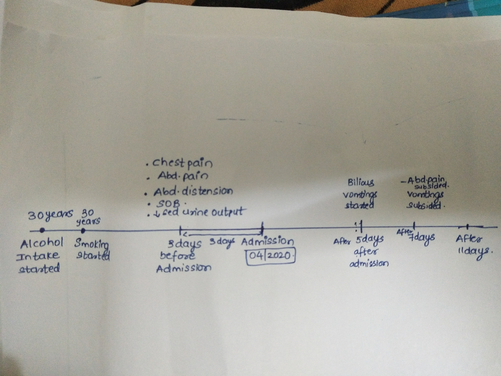
Case2
54 year old male with cough,abdominal tightness,pedal edema and diarrhea.
When exposed to bactericidal concentrations of antimicrobial drugs, the number of viable cells in a susceptible bacterial population does not decline exponentially. Instead, the mortality rate decreases over time and a substantial fraction of the population may survive antimicrobial drug treatment. This phenomenon has been observed for virtually all antimicrobials used in clinical practice and for many bacterial species and has been attributed to antimicrobial tolerance. Phenotypic antimicrobial tolerance is a temporary, reversible bacterial state that is often associated with a reduced rate of multiplication, where some antimicrobial drugs are ineffective against genetically susceptible bacilli. Antimicrobial tolerance is hypothesised to be the prime reason for the extended treatment regimens required for M.tb chemotherapy, as fully drug-sensitive bacilli survive (persist through) initial antimicrobial drug therapy. It has long been speculated that M.tb in a non/slowly-replicating state may play a clinically significant role, persisting during drug therapy.The first-line antimicrobials used to treat M.tb infection (isoniazid, rifampicin, pyrazinamide and ethambutol) are all active against actively-replicating bacteria, however effectiveness of this drug regimen against non/slowly-replicating bacilli is reduced or eliminated. This M.tb slow/non-replicating state is hypothesised to be induced by the environmental conditions found in specific granuloma types, in particular those associated with hypoxia or nitric oxide production.An M.tb transcriptional signature resembling slow or non-replicating bacilli was also identified in bacilli isolated from human sputa. Exposure of bacilli to such microenvironments results in the expression of a discrete set of genes known as the dormancy regulon (DosR/DevR) that are in turn responsible for transitioning bacilli into a non-replicating and hence likely drug-tolerant state. The mechanism(s) that result in the generation of phenotypic drug tolerant M.tb populations in vivo are currently not well understood, however it is critical to consider these sub-populations of bacilli for the development of more effective drug regimes.
The M.tb population in vivo has been compared to a Russian nesting doll, consisting of layer after layer of distinct bacterial subpopulations that may also be separated by time and space, each of which may be differentially killed by various antimicrobial drugs dependent on phenotypic drug tolerance or anatomical location. The models developed by Mitchison and Mitchison and Coates are often adapted to describe four populations defined by antimicrobial drug efficacy (1) actively growing bacilli mostly killed by isoniazid, (2) slow/non-replicating M.tb bacteria that undergo spurts of metabolism, which are killed by rifampicin, (3) intracellular bacilli present in the acidic compartments of macrophages or in acidic lung lesions that are killed by pyrazinamide, and (4) M.tb persisters found in hypoxic microenvironments with much reduced action of most anti-TB drugs. As evidenced in TB patients that relapse during treatment of drug-sensitive M.tb, the host immune system cannot effectively eliminate these residual M.tb bacilli that are not killed by chemotherapy. Therefore, although achieving a clinical cure, the current anti-TB standard regimen does not necessarily achieve a bacteriological cure. In other words, current therapy does not completely eradicate all bacilli from the body, but allows the infection to be contained effectively for long-periods of time. These observations underscore the need for developing better sterilising compounds against M.tb. However, the lack of adequate screening systems able to identify new compounds effective against drug-tolerant M.tb phenotypes remains an immense barrier to the anti-tuberculosis drug development process.
*Spot urine protein-to-creatinine ratio can be used instead of 24-hour urine collection to confirm nephrotic-range proteinuria. |
Metabolic | |
Amyloidosis | |
Diabetes mellitus | |
Immunologic | |
Cryoglobulinemia | |
Erythema multiforme | |
Henoch-Schönlein purpura | |
Microscopic polyangiitis | |
Polyarteritis nodosa | |
Sjögren syndrome B) Renal biopsy Pros of renal biopsy: In patients with NS from a known secondary cause and who are responding to treatment appropriately, biopsy will likely add little to treatment. Biopsy may be more useful for treatment and prognosis in patients with idiopathic NS of an unknown histologic disease type or with suspected underlying systemic lupus erythematosus or other renal disorders. Cons of renal biopsy:
|